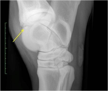
Equine Osteochondrosis: A controversial disease with multifactorial etiology
Keywords:
equine, osteochondrosis, growth cartilage, subchondral bone, multifactorial etiologyAbstract
Osteochondrosis (OC) is an osteoarticular disorder that mainly affects humans, pigs, horses and dogs. Is one of the most common joint diseases of growing horses, belonging to developmental orthopedic diseases complex. It is characterized by an alteration of the endochondral ossification that affects growth of cartilage and the subchondral bone, and is considered the primary phase of damage to joint-epiphyseal complex. Its etiology is reported to be multifactorial and etiological factors most often described are heredity, dietary and endocrine imbalances, rapid growth, trauma and vascular defects of the growth cartilage, but it is incompletely understood, and there is great controversy about the participation and interaction of these factors in appearance of the disease. Osteochondrosis is a disease that causes great economics losses in horse industry, representing an important problem for the sector, hence the great interest in determining its origin.
Downloads
References
[2] Bohndorf K. Osteochondritis (osteochondrosis) dissecans: a review and new MRI classification. Eur Radiol 1998; 8:103-112.
[3] Distl O. The genetic of equine osteochondrosis. Vet J 2013; 197(1):13-18.
[4] McIlwraith CW. Diseases of joints, tendons, ligaments, and related structures. En: Stashak TS, editor. Adams’ Lameness in horses. 5 ed. Lippincott Williams & Wilkins, Baltimore, USA; 2002. p. 459-644.
[5] Jans L, Jaremko J, Ditchfield M, Verstraete K. Evolution of femoral condylar ossification at MR imaging: frequency and patient age distribution. Radiology 2011; 258(3):880-888.
[6] van Weeren PR. Natural history of and recommendations for OC lesions. Proceedings of the 13th ESVOT Congress. 2006, Sep. 7-10; Munich, Germany.
[7] van Weeren PR. Etiology, diagnosis, and treatment of OC(D). Clin Tech Equine Pract 2006; 5:248-258.
[8] Douglas J. Pathogenesis of osteochondrosis. En: Ross MW y Dyson SJ, editores. Lameness in the Horse. Elsevier Science, USA; 2003. p. 534-543.
[9] Watkins JP. Osteochondrosis. En: Auer, J, editor. Equine Surgery. WB Saunders, USA; 1992. p. 971-984.
[10] Barrie HJ. Osteochondritis dissecans 1887-1987. A centenal look at König’s memorable phrase. J Bone Joint Surg Br 1987; 69:693-695.
[11] Nilsson F. Hästens goniter. Sven Vet Tidskr 1947; 52:1-14.
[12] Birkeland, R. Chip fractures of the first phalanx in the metatarsophalangeal joint of the horse. Acta Radiol Suppl 1972; 319:73-77.
[13] De Moore A, Verschooten F, Desmet P, Steenhaut M, Hoorens J, Wolf G. Osteochondritis dissecans of the tibiotarsal joint of the horse. Equine Vet J 1972; 4(3):139-143.
[14] Poulos P. Radiologic manifestations of developmental orthopedic disease. Proceedings of AQHA Developmental Orthopedic Disease Symposium. 1986, April 21-22; Dallas, USA.
[15] Desjardin C, Riviere J, Vaiman A, Morgenthaler C, Diribarne M, Zivy M, et al. Omics technologies provide new insights into the molecular physiopathology of equine osteochondrosis. BMC Genomics 2014; 15(1): 947-958.
[16] Dik KJ, Enzerink E, van Weeren PR. Radiographic development of osteochondral abnormalities, in the hock and stifle of Dutch Warmblood foals, from age 1 to 11 months. Equine Vet J 1999; Supl (31):9-15.
[17] Wittwer C, Hamann H, Rosenberger E, Distl O. Prevalence of osteochondrosis in the limb joints of South German Coldblood horses. J Vet Med A Physiol Pathol Clin Med 2006; 53(10):531-539.
[18] Oliver LJ, Baird DK, Baird AN, Moore GE. Prevalence and distribution of radiographically evident lesions on repository films in the hock and stifle joints of yearling Thoroughbred horses in New Zealand. NZ Vet J 2008; 56(5):202-209.
[19] Jönsson L, Dalin G, Egenvall A, Näsholm A, Roepstorff L, Philipsson J. Equine hospital data as a source for study of prevalence and heritability of osteochondrosis and palmar/plantar osseous fragments of Swedish Warmblood horses. Equine Vet J 2011; 43(6):695-700.
[20] Boado A, López-Sanromán FJ. Prevalence and characteristics of osteochondrosis in 309 Spanish Purebred horses. Vet J 2016; 207:112-117.
[21] Lim CK, Hawkins JF, Vanderpool AL, Geng HG, Harmon CCG, Lenz SD. Osteochondritis dissecans-like lesions of the occipital condyle and cervical articular process joints in a Saddlebred colt horse. Acta Vet Scand 2017; 59:76-82.
[22] Richardson DC, Zentek J. Nutrition and osteochondrosis. Vet Clin North Am Small Anim Pract 1998; 28:115-135.
[23] Bridges CH, Womack JE, Harris ED, Scrutchfield WL. Considerations of cooper metabolism in osteochondrosis of suckling foals. J Am Vet Med Ass 1984; 185(2):173-178.
[24] Gee E, Davies M, Firth E, Jeffcott L, Fennessy P. Osteochondrosis and cooper: histology of articular cartilage from foals out of cooper supplemented and non-supplemented dams. Vet J 2007; 173:109-117.
[25] van Weeren PR, Knaap J, Firth EC. Influence of liver cooper status of mare and newborn foal on the development of osteochondrosis lesions. Equine Vet J 2003; 35(1):67-71.
[26] Kowalczyk DF, Gunson DE, Shoop CR, Ramberg CFJr. The effects of natural exposure to high levels of zinc and cadmium in the immature pony as a function of age. Environ Res 1986; 40(2):285-300.
[27] Gunson D, Kowalczyk D, Shoop CR, Ramberg CJr. Environmental zinc and cadmium pollution associated with generalized osteochondrosis, osteoporosis, and nephrocalcinosis in horses. J Am Vet Assoc 1982; 180(3):295-299.
[28] Campbell-Beggs CL, Johnson PJ, Messer NT, Lattimer JC, Johnson G, Casteel SW. Osteochondritis dissecans in an Appaloosa foal associated with zinc toxicosis. J Equine Vet Sci 1994; 14(10):546-550.
[29] Swerczek TW. Chronic environmental cadmium toxicosis in horses and cattle. J Am Med Assoc 1997; 211(10):1229-1230.
[30] Savage CJ, McCarthy RN, Jeffcott LB. Effects of dietary phosphorus and calcium on induction of dyschondroplasia in foals. Equine Vet J 1993; 25 Supl 16:80-83.
[31] Reiland S. Effects of vitamin D and A, calcium, phosphorus, and protein on frequency and severity of osteochondrosis in pigs. Acta Radiol 1978; 358 Supl:91-105.
[32] Bruns J, Werner M, Soyka M. Is vitamin D insufficiency or deficiency related to the development of osteochondritis dissecans? Knee Surg Sports Traumatol Arthrosc 2016; 24(5):1575-1579.
[33] Vander Heyden L, Lejeune JP, Caudron I, Detilleux J, Sandersen C, Chavatte P, et al. Association of breeding conditions with prevalence of osteochondrosis in foals. Vet Rec 2013; 172(3):68-71.
[34] Jeffcott LB, Henson FM. Studies on growth cartilage in the horse and their application to aetiopathogenesis of dyschondroplasia (osteochondrosis). Vet J 1998; 156:177-192.
[35] Pagan JD, Geor RJ, Caddel SE, Pryor PB, Hoekstra KE. The relationship between glycemic response and the incidence of OCD in thoroughbred weanlings: a field study. 47th Proceedings of the Annual Convention of the AAEP. 2001, Nov. 24-28; San Diego, U.S.A.
[36] Kronfeld DS, Meacham TN. Dietary aspects of developmental orthopedic disease in young horses. Vet Clin North Am Equine Pract 1990; 6(2):451-465.
[37] Strömberg B. A review of the salient features of osteochondrosis in the horse. Equine Vet J 1979; 11(4):211-214.
[38] Donabédian M, Fleurance G, Robert C, Lepage O, Leger S, et al.. Effect of fast vs. moderate growth rate related to nutrient intake on developmental orthopaedic disease in the horse. Anim Res 2006; 55 (5):471-486.
[39] Gamboa A, Garzón-Alvarado DA. Factores mecánicos en enfermedades osteocondrales. Rev Cubana Invest Bioméd 2011; 30(1):174-193.
[40] de Koning DB, van Grevenhof EM, Laurenssen BF, Ducro BJ, Heuven HC, de Groot PN, et al.. Associations between osteochondrosis and conformation and locomotive characteristics in pigs. J Anim Sci 2012; 90(13):4752-4763.
[41] Hernández G, Rodríguez L, Mora F, Ramírez R. Etiología, patogénesis, diagnóstico y tratamiento de osteocondrosis (OC). Vet Méx 2011; 42(4):311-329.
[42] McCoy AM, Toth F, Dolvik NI, Ekman S, Ellermann J, Olstad K, et al. Articular osteochondrosis: a comparison of naturally-occurring human and animal disease. Osteoarthritis Cartilage 2013; 21(11):1638-1647.
[43] Lepeule J, Bareille N, Robert C, Valette JP, Jacquet S, Blanchard G, et al. Association of growth, feeding practices and exercise conditions with the severity of the osteoarticular status of limbs in French foals. Vet J 2013; 197(1):65-71.
[44] Lewczuk D, Korwin-Kossakowska A. Genetic background of osteochondrosis in the horse – a review. Anim Sci Pap Rep 2012; 30(3):205-218.
[45] van Weeren, PR, Olstad K. Pathogenesis of osteochondrosis dissecans: How does this translate to management of the clinical case? Equine Vet Educ 2016; 28(3):155-166.
[46] Atanda A, Shah S, O`Brien K. Osteochondrosis: common causes of pain in growing bones. Am Fam Physician 2011; 83(3):285-291.
[47] Reiland S, Ordell N, Lundeheim N, Olsson SE. Heredity of osteochondrosis, body constitution and leg weakness in the pig. A correlative investigation using progeny testing. Acta Radiol 1978; Supl 358:123-137.
[48] LaFond E, Breur GJ, Austin CC. Breed susceptibility for developmental orthopedic disease in dogs. J Am Anim Hosp Assoc 2002; 38:467-477.
[49] Grøndahl AM, Dolvik NI. Heritability estimations of osteochondrosis in the tibiotarsal joint and of bony fragments in the palmar/plantar portion of the metacarpo- and metatarsophalangeal joints of horses. J Am Vet Med Assoc 1993; 203:101-104.
[50] Russell J, Matika O, Russell T, Reardon RJ. Heritability and prevalence of selected osteochondrosis lesions in yearling Thoroughbred horses. Equine Vet J 2017; 49(3):282-287.
[51] Philipsson J. Pathogenesis of osteochondrosis – genetic implications. En: McIlwraith CW y Trotter GW, editores. Joint Disease in the Horse. WB Saunders, USA; 1996. p. 359-362.
[52] Schougaard H, Falk J, Phillipson, J. A Radiographic survey of tibiotarsal osteochondrosis in a selected population of trotting horses in Denmark and its possible genetic significance. Equine Vet J 1990; 22(4):288-289.
[53] Pieramati C, Pepe M, Silvestrelli M, Bolla A. Heritability estimation of osteochondrosis dissecans in Maremmano horses. Livest Prod Sci 2003; 79(2-3):249-255.
[54] Carlson CS, Cullins LD, Meuten JD. Osteochondrosis of the articular-epiphyseal cartilage complex in young horses: evidence for a defect in cartilage canal blood supply. Vet Pathol 1995; 32:641-647.
[55] Olstad K, Ytrehus B, Ekman S, Carlson CS, Dolvik NI. Early lesions of osteochondrosis in the distal femur of foals. Vet Pathol 2011; 48:1165-1175.
[56] Olstad K, Østevik L, Carlson CS, Ekman, S. Osteochondrosis can lead to formation of pseudocysts and true cysts in the subchondral bone of horses. Vet Pathol 2015; 52(5):862-872.
[57] Ytrehus B, Haga HA, Mellum CN, Mathisen L, Carlson CS, Ekman S, et al. Experimental ischemia of porcine growth cartilage produce lesions of osteochondrosis. J Orthop Res 2004; 22(6):1201-1209.
[58] Olstad K, Ytrehus B, Ekman S, Carlson CS, Dolvik NI. Epiphyseal cartilage canal blood supply to the tarsus of foals and relationship to osteochondrosis. Equine Vet J 2008; 40(1):30-39.
[59] Laverty S, Girard C. Pathogenesis of epyphiseal osteochondrosis. Vet J 2013; 197(1):3-12.
[60] van Weeren, PR. Equine Osteochondrosis: a challenging enigma. Pferdeheilkunde 2005; 21(4):285-292.
[61] Edmonds EW, Polousky J. A review of knowledge in osteochondritis dissecans: 123 years of minimal evolution from König to the ROCK study group. Clin Orthop Relat Res 2013; 471:1118–1126.

Published
How to Cite
Issue
Section
Gaceta de Ciencias Veterinarias se apega al modelo Open Access, por ello no se exige suscripción, registro o tarifa de acceso a los usuarios o instituciones. Los usuarios pueden leer, descargar, copiar, distribuir, imprimir y compartir los textos completos inmediatamente después de publicados, se exige no hacer uso comercial de las publicaciones. Para la reproducción parcial o total de los trabajos o contenidos publicados, se exige reconocer los derechos intelectuales de los autores y además, hacer referencia a esta revista. La publicación de artículos se hace sin cargo para los autores. Los trabajos pueden consultarse y descargarse libremente, y de manera gratuita, en extenso en versión digital, desde su enlace Web institucional. Los textos publicados son propiedad intelectual de sus autores. Las ideas, opiniones y conceptos expuestos en los trabajos publicados en la revista representan la opinión de sus autores, por lo tanto, son estos los responsables exclusivos de los mismos.


